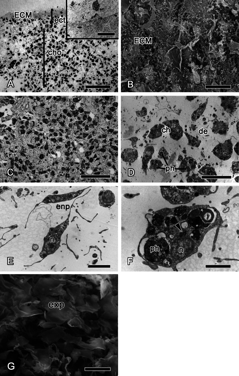
Figure 4. 12 h after injury.
(A) Semi thin section of wounded ectosome and adjacent area of choanosome. Inset: middle layer of ectosome, containing collagen fibrils organized into firm tracts. (B) SEM of wound surface, covered with ECM. (C) Semi thin section of wounded choanosome. (D) TEM of dedifferentiated cells in wounded choanosome. (E) TEM of dedifferentiated endopinacocytes in wounded choanosome. (F) TEM of dedifferentiated choanocyte filled with phagosomes, but with remaining basal body and accessory centriole (arrowhead). (G) SEM of intact exopinacocytes, surrounding the wound. ch, choanocytes; cho, choanosome; de, debris; ect, ectosome; ECM, extra cellular matrix; enp, endopinacocytes; exp, exopinacocytes; n, nucleus; ph, phagosomes. Scale bars: A—50 µm, B—10 µm; C—25 µm; D—15 µm; E—4 µm; F—2 µm; G—10 µm.

