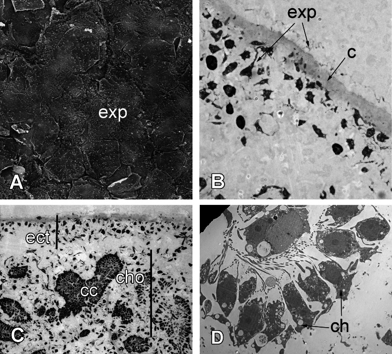
Figure 8. 72 h after injury.
(A) SEM of the new exopinacoderm. (B) TEM of regenerated ectosome. (C) Semi-thin section of regenerated ectosome (ect) and choanosome (cho). (D) TEM of new choanocyte chamber. Scale bars: A—20 µm; B—20 µm; C—50 µm; D—5 µm. c, cortex layer; cc, choanocyte chamber; ch, choanocytes; cho, choanosome; ect, ectosome; exp, sirface of new exopinacocytes.

