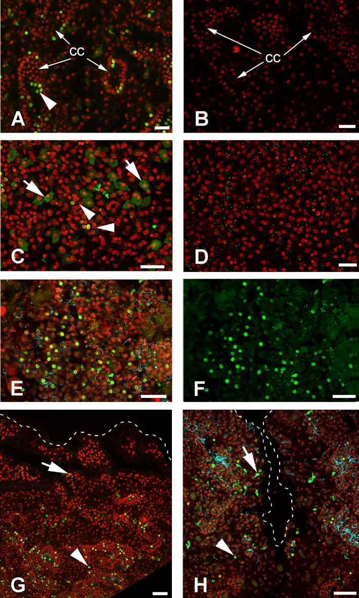
Figure 9. DNA synthesis in unwounded Halisarca dujardini and during regeneration.
(A) DNA synthesis in choanocytes of unwounded sponge after 6 h incubation with EdU. (B) Negative control for A sample, incubated 6 h without EdU. (C) EdU incorporation in nuclei (arrowhead) and cytoplasm of cells after 24 h incubation. (D) Negative control for C sample, incubated 24 h without EdU. (E), (F) Wound surface after 12 h of regeneration. (F) Green channel (EdU) only. (G) Transversal section of wound surface at 24 h of regeneration. (H) Sagittal section of wound surface at 24 h of regeneration at peripheral level. Wound border outlined with dashed line. Cyan—tubulin, Red—DNA, green—EdU. cc, choanocyte chamber. Arrowheads indicate labeled nuclei, arrows—labeled cytoplasmic granules of unknown nature. Scale bars: A–D—10 µm; E, F—20 µm; G, H—30 µm.

