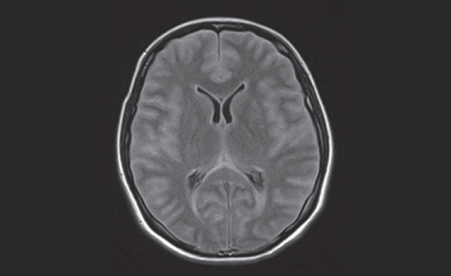
Figure 3).

Magnetic resonance examination of the brain revealing diffuse sulcal space and cisternal space effacement with diffusely increased signal of the extra-axial cerebrospinal fluid spaces and ependyma of the lateral ventricles on fluid-attenuated inversion recovery imaging. Diffuse parenchymal swelling and slightly increased T2-weighted signal of the cortex of the temporal lobes, insular cortex and hippocampal regions noted bilaterally suggests encephalitis
