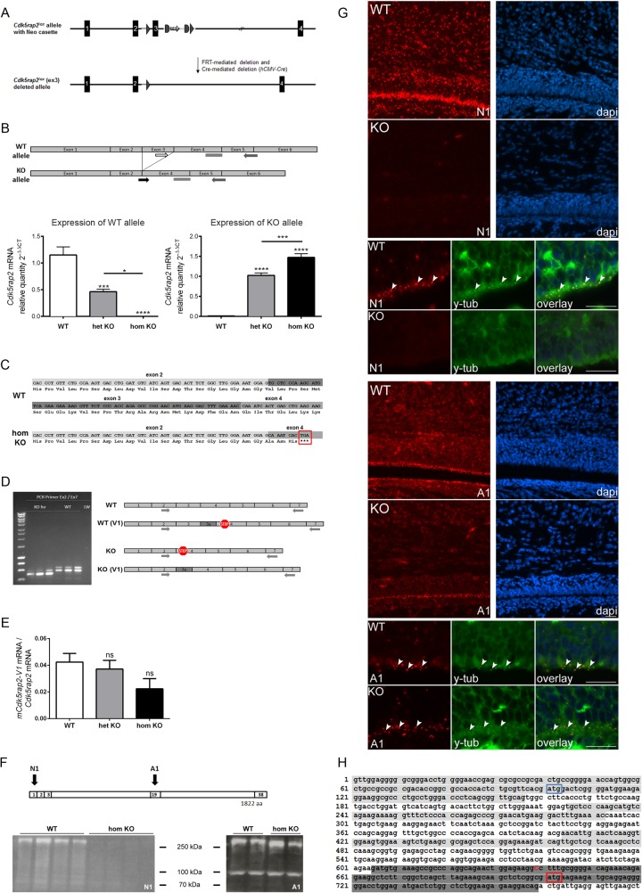
Fig 1. Conditional Cdk5rap2 KO mouse and novel mCdk5rap2 splice variants.
(A) Cdk5rap2 lox construct before and after Cre-mediated deletion of exon 3. (B) Cdk5rap2 WT and KO allele expression in the neocortex of WT, het KO, and hom KO mice at P0. The WT allele is not expressed in hom KO mice; the KO allele is not expressed in WT mice (qPCR, n = 6 per group, one-way ANOVA, p<0.0001; Bonferroni’s Multiple Comparison Test). Position of forward primers specific for the WT and the KO allele, respectively, and the common reverse primer and probe are depicted. (C) Sanger sequencing results of PCR products from exon 2 to exon 7 of WT and hom KO cDNA confirmed the correct excision of exon 3 in hom KO mice, resulting in a frame shift and a premature stop codon (red box). (D) Identification of a novel mCdk5rap2 splice variant (mCdk5rap2-V1) through gel electrophoresis and sequencing of PCR products from exon 2 to exon 7 of WT and hom KO cDNA. In hom KO mice, the additional 71 nucleotides will abolish the frameshift and stop codon introduced through excision of exon 3. In WT mice the additional 71 nucleotides will lead to a frameshift and a premature stop codon resulting in a truncated 85 aa protein. (E) mCdk5rap2-V1 mRNA expression in the neocortex of WT, het KO, and hom KO mice at P0 (qPCR, n = 6 per group, one-way ANOVA, p = 0.1314; Bonferroni’s Multiple Comparison Test). (F) Western blot analysis using the N1 antibody revealed a strong reduction of protein levels below detection levels in hom KO in comparison to WT neocortex at P0, while multiple bands were identified with a reduction in the size of the largest (about 250 kDa) band by about 10 kDa when using the A1 antibody. Binding sites of anti-Cdk5rap2 antibodies N1 and A1 are depicted, numbers refer to exons encoding the corresponding protein regions. (G) Immunostaining using antibodies directed against Cdk5rap2 (red) and the centrosome marker y-tubulin (green) of coronal murine hom KO and WT brain sections at P0; nuclei are stained with DAPI (blue). Overview immunofluorescence pictures and higher magnification images of the subventricular (SVZ) and the ventricular (VZ) zone; arrowheads indicate examples for centrosomes which are co-stained with Cdk5rap2 and y-tubulin; scale bars 20 μm. When applying the N1 antibody, high Cdk5rap2 signal density is present within the neocortex and SVZ/VZ in WT mice, while this is lacking in hom KO mouse brains. The A1 antibody, however, produces Cdk5rap2 immunopositivity in both the hom KO and WT neocortex and SVZ/VZ with a high signal density in the in the WT mice and a similar pattern with only a slight decrease of staining density and intensity in hom KO mice. (H) Excerpt of Cdk5rap2 mRNA sequence (NM_145990.3, exons 1, 3, and 5 are highlighted in light grey, exon 7 in dark grey) with known start codon in exon 1 (blue box) and alternative start codon in exon 7 (red box) as well as putative sequence start of shorter variant Cdk5rap2-V2 (red upper case C). ns, not significant, *p<0.05, **p<0.01, ***p<0.001, ****p<0.0001.

