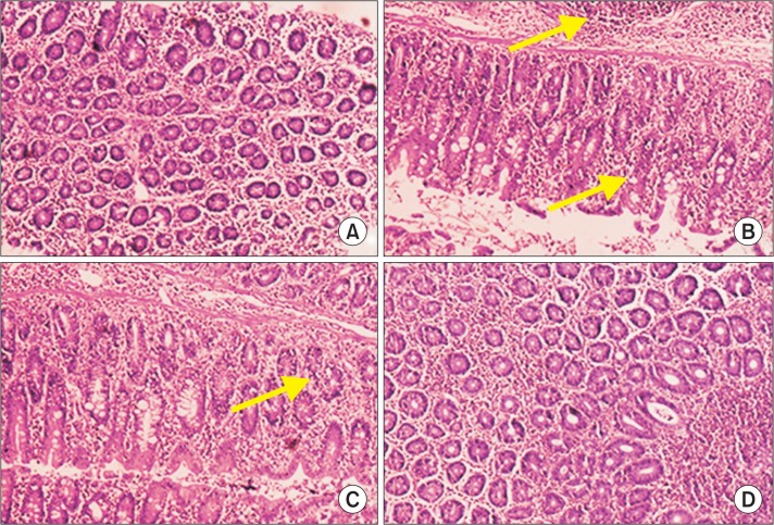
Fig. 4.
Histopathological analysis of control and experimental animals. (A) shows normal architecture of control animals with effective staining in nucleus and cytoplasm. (B) express distorted nuclei and enlarged size of nuclei with closely packed glands causing infiltration of cancer cells with invasive adenoma. Villus and goblet cells of simple coloumnar epithelium show profound staining with reference to cellular damage and diffusion into lymphatic system. (C) show regeneration of villus and epithelial linings with respect to myrtenal treatment and decreased infiltration of cancer cells into the connective tissue. (D) show normal morphology of experimental animals with myrtenal supplementation alone showing no toxicity by cellular damage.

