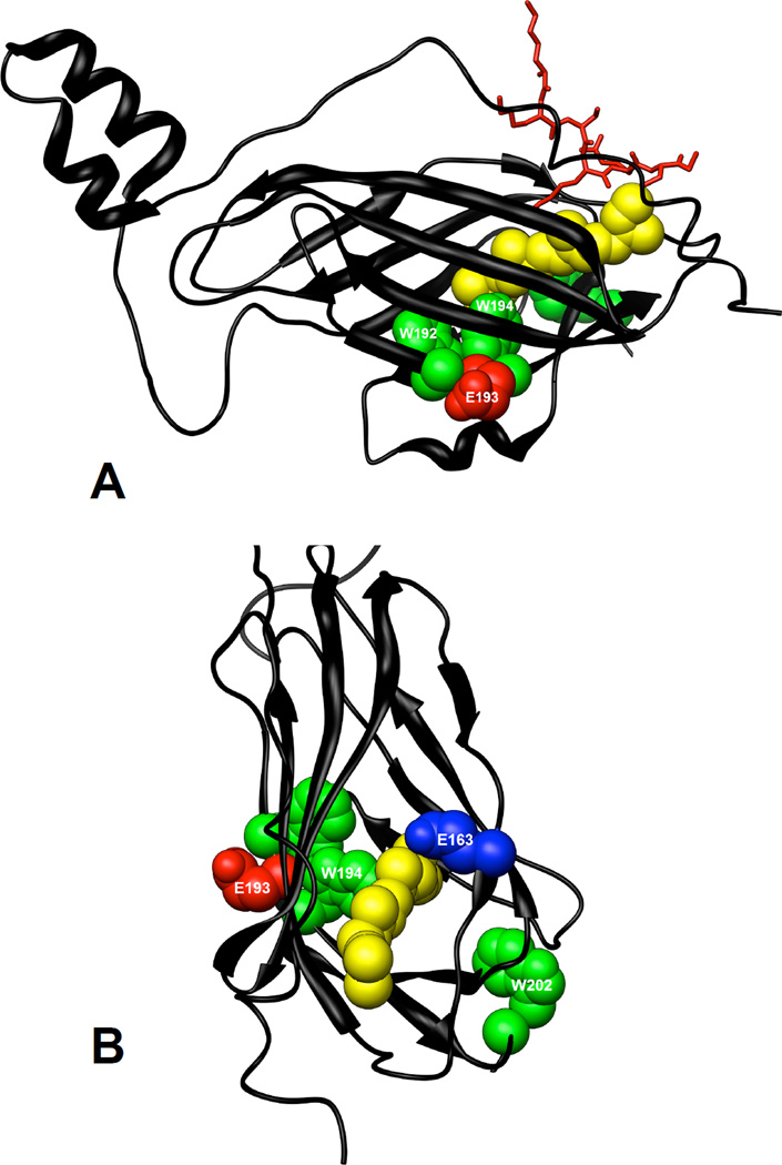
Figure 11.
Location of tryptophans and residues photolabeled by 3-azibutanol within the structure of RhoGDIα. The crystal structure of RhoGDIα in complex with Cdc42 (not shown) was from Hoffman et al (16). Tryptophans, W192, W194 and W202 are shown in green, residues photolabeled by 3-azibutanol are shown in red and the GG chain of Cdc42 is shown in yellow. The C-terminal backbone of Cdc42 (K183-C188) is shown in red. The Protein Data Bank file number of the structure used is 1DOA and the molecular graphics images were produced using the UCSF Chimera package (36)

