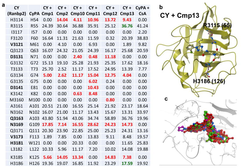
FIGURE 7. Structural poses of CY-ligand complexes.
(a) Surface contact areas (Å2) between docked ligands of CY and residues of PPIase pocket of CY determined by molecular modeling with ICM. Counterpart residues in CyPA are also shown (2nd column). Non-conserved residues in CY are shown in bold. Contact areas (Å2) between docked CsA and residues of PPIase pocket of CyPA as determined by X-ray crystallography are shown (last column). Numbers in red denote residues establishing interactions with chemical probes identified by this study and that are known not to participate in interactions between CyPA and CsA. Residues in bold are unique to CY of Ranbp2. Residues in green establish hydrogen bonds with compound 13. (b) Hydrogen bonding between compound 13 and the conserved residues, R3115 and H3186, of the PPIase pocket of CY. Numbers in parenthesis are counterpart residues in CyPA. (c) Superposition of ligands of PPIase pocket of CY; compound 11 (purple) protrudes out the PPIase pocket of CY.

