
Fig. 1.
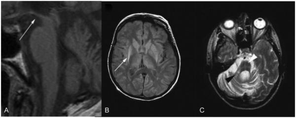
Structural magnetic resonance imaging in progressive supranuclear palsy (PSP) and multiple system atrophy (MSA). (A) T1-weighted image showing hummingbird sign (white arrow) in PSP 1.5 years after onset. (B) Proton density weighted image of pathologically confirmed MSA. Putaminal atrophy with hyperintense putaminal rim (white arrow) on the right and early hyperintense putaminal rim on the left 4.8 years after disease onset. (C) Right middle cerebellar peduncle sign (black arrowhead) and hot cross bun sign (white arrowhead) in pathologically confirmed MSA (T2-weighted image). (Adapted with permission from Massey et al. Conventional Magnetic Resonance Imaging in Confirmed Progressive Supranuclear Palsy and Multiple System Atrophy. Mov Disord 2012; 27:1754–1762.)