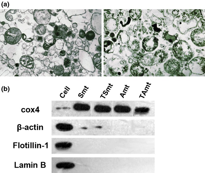
Figure 2.

(a) Electron microscopy image of mitochondria morphology (10 000×). The purity of the mitochondria was confirmed using electron microscopy, including intactness and contamination. (b) Western blot analyses of total cell proteins (Cell) and mitochondrial preparations isolated from paclitaxel-sensitive SKOV3 cells (Smt), paclitaxel-resistant SKOV3 cells (TSmt), paclitaxel-sensitive A2780 cells (Amt), and paclitaxel-resistant A2780 cells (TAmt) for COX4 (mitochondrial marker), lamin-B (nuclear marker), flotillin-1 (cell membrane marker) and β-actin (cytoskeletal protein). There was little contamination in the mitochondria-enriched fractions.
