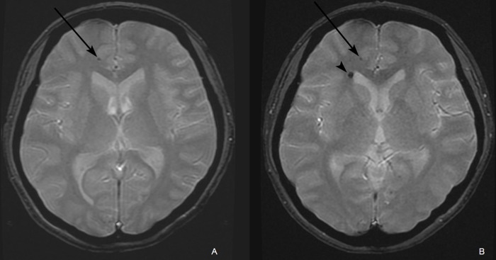
Fig 1. A of a 34-year-old female who received radiation therapy due to an ependymoma in the fourth ventricle.
(A) A gradient echo (GRE) image acquired five years following radiation therapy revealed a small dark dot lesion (arrow) in the subcortical white matter of the right frontal lobe. (B) A GRE image acquired seven years following radiation therapy revealed a newly developed intracerebral hemorrhage (arrowhead) adjacent to the right lateral ventricle frontal horn next to the previously noted intracerebral hemorrhage in the subcortical white matter of right frontal lobe (arrow).

