Figure 3.
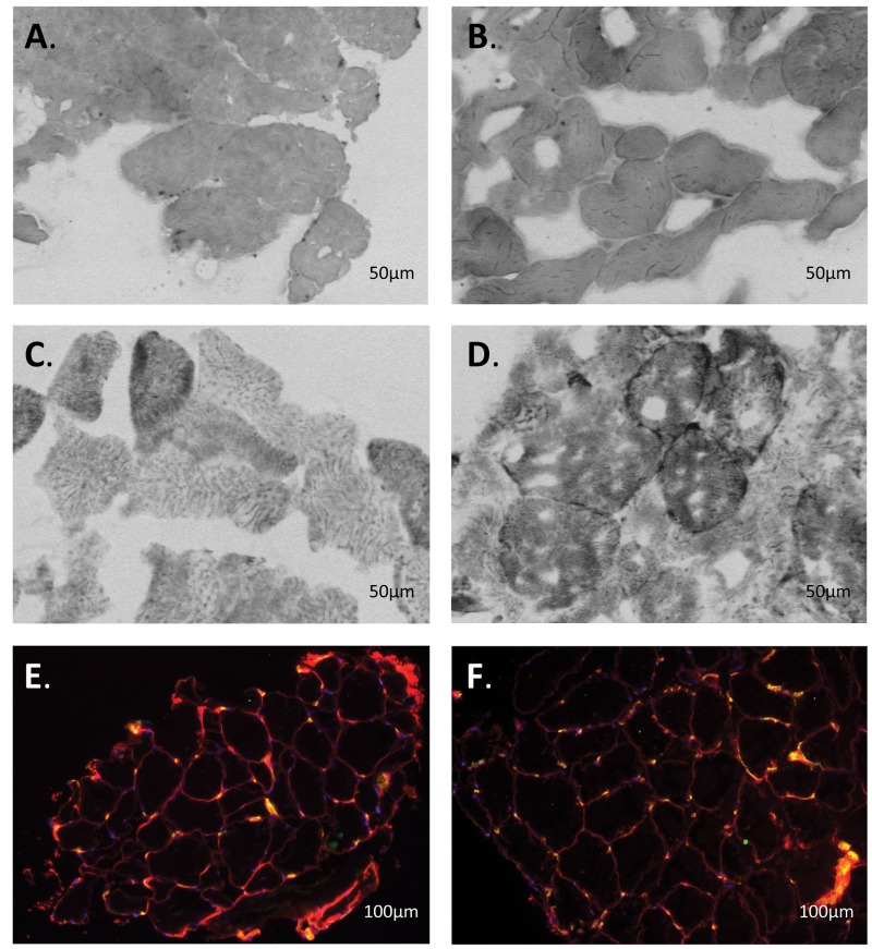
Quadriceps biopsy samples from idiopathic pulmonary arterial hypertension (iPAH) patients. Shown are typical examples of quadriceps biopsy samples from iPAH patients before (A, C, E) and after (B, D, F) intravenous iron therapy. A, B, Myoglobin staining. C, D, Succinate dehydrogenase activity staining. Type I and II cells are distinguished by color, as type I cells have more myoglobin and succinate dehydrogenase activity (darker cells) than type II cells. E, F, CD31 staining. Every yellow dot represents a capillary.

