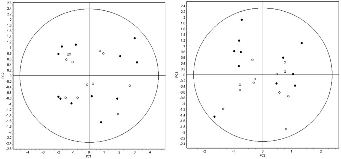
Figure 2.

Principal-component analysis of the 24 pooled samples (12 left ventricular [LV], 12 right ventricular [RV]) using 407 features that were matched across all of the 12 gels. LV samples are represented by filled circles, and RV samples are represented by open circles. Both groups encompass explanted hearts from patients with or without ischemic and/or RV echocardiographic dysfunction. The first 3 principal components cumulatively represent more than 50% of the variation in the analysis (PC1: 37.4%, PC2: 10.3%, PC3: 9.8%) yet fail to segregate the samples into any discrete groups based on either biology of unanticipated technical variation. Thus, the magnitude and/or the number of statistically significant changes in protein expression between the right and left ventricles is expected to be low.
