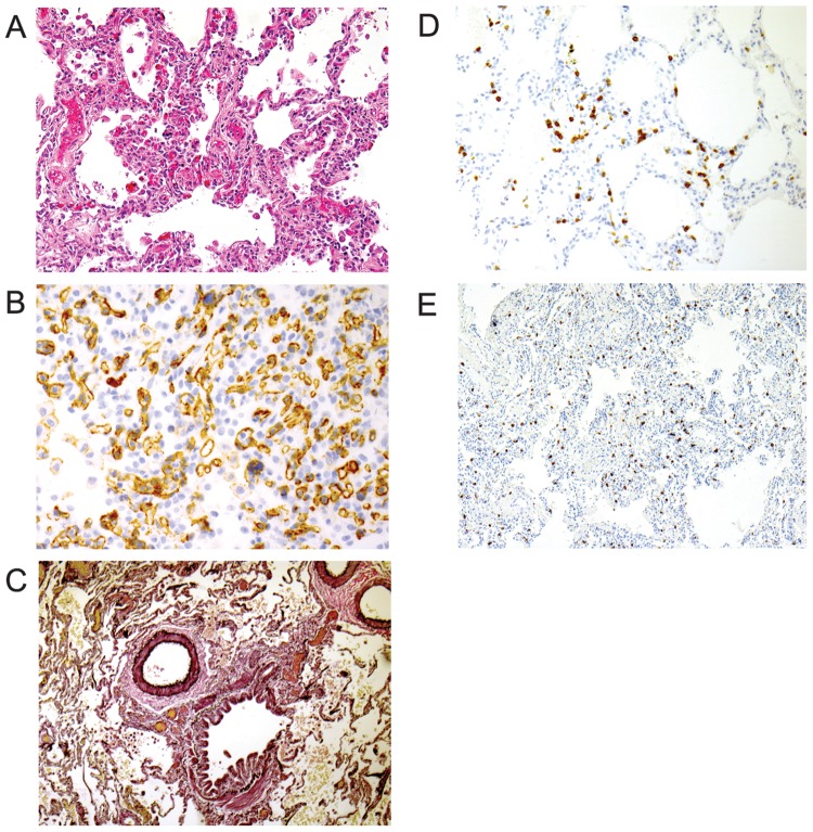
Figure 1.
A, Lung shows focal proliferation and duplication of alveolar capillaries with thickened walls and dilated lumens that are characteristic of pulmonary capillary hemangiomatosis. B, CD31 immunostain highlights the proliferating endothelial cells. C, An elastic stain shows normal-caliber pulmonary artery adjacent to a bronchiole. D, CD3 immunostain highlights the T cell alveolitis. E, An immunostain for tryptase highlights the expanded interstitial mast cell population.

