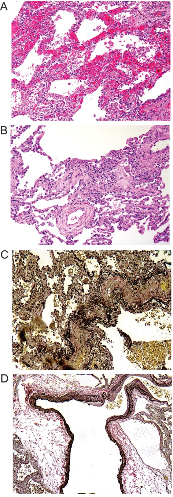
Figure 3.

A, Lung shows the features of pulmonary capillary hemangiomatosis. B, Interstitial lymphocytic alveolitis is seen in a hematoxylin and eosin–stained section. C, Veno-occlusive changes of small venules are demonstrated by elastic stain. D, An arteriovenous malformation is highlighted by an elastic stain.
