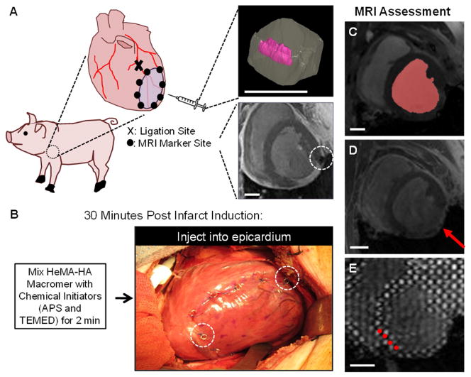
Fig. 2.
MRI analysis of in vivo porcine infarct model. In vivo function (n=3–6/group) was assessed in an established porcine posterior infarct model (A). Inserts in panel A show a three-dimensional MRI reconstruction of a hydrogel injection in a myocardial explant (top) and visibility of a MRI compatible marker (white, dashed circle) placed post-MI for tracking infarct expansion over time (bottom). Thirty minutes post-MI, animals underwent an array of twenty 0.3 mL injections of either prepolymer solution or saline in the infarct (B). MRI compatible markers (white, dashed circles) are visible. MRI scans were performed at baseline (i.e. prior to infarction) and at 1, 4, 8 and 12 weeks post-MI. MRI data was analyzed to assess global LV structure and function from segmentation of the blood volume (red shape) in cine MRI (C), infarct (red arrow) expansion from LGE MRI (D), and myocardial wall function from tracking grid displacement (red circles highlight select tags) in SPAMM tagged MRI (E). Scale bar = 2 cm.

