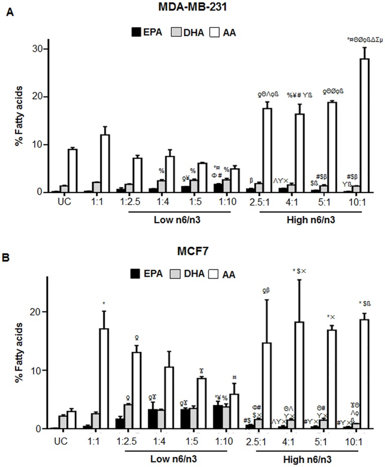
Fig 8. Relative percentage of EPA, DHA and AA in breast cancer cell lines.
The cells were treated with different ratios of n6 and n3 FA for 24h. The levels of EPA, DHA and AA has been shown in MDA-MB-231 (A) and MCF7 (B) cells treated with low and high n6/n3 ratios. Each value represents mean±SEM of three independent experiments. %p<0.05, ƍp<0.01 and *p<0.001 compared to UC; Ϫp<0.05, ¥p<0.01 and ¤p<0.001 compared to 1:1; Φp<0.01 and Θp<0.001 compared to 1:2.5; #p<0.05, Λp<0.01 and Øp<0.001 compared to 1:4; $p<0.05, ϒp<0.01 and ϙp<0.001 compared to 1:5; βp<0.05, ×p<0.001 and ßp<0.001 compared to 1:10; Δp<0.001 compared to 2.5:1; Σp<0.001 compared to 4:1; μp<0.01 compared to 5:1.

