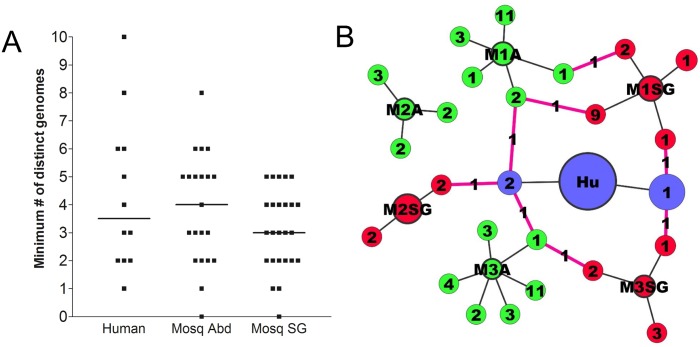
Fig 7. Phasing SNVs into distinct variant viral genomes.
(A) Minimum numbers of distinct variant DENV genomes per sample, in human- mosquito abdomen-, and mosquito salivary gland-derived DENV2 populations. (B) Transmission network for patient-mosquito group 641. For each sample, consensus viral genomes are represented by circles outlined in bold (Hu, human; M1A, M2A, M3A, mosquito abdomens; M1SG, M2SG, M3SG, mosquito salivary glands), and variant genomes by circles radiating out of the consensus circle. Numbers within variant circles indicate the number of SNVs present in each variant genome. Pink connector lines represent maintenance of SNV across transmission stages, with numbers indicating the number of SNVs that were maintained.

