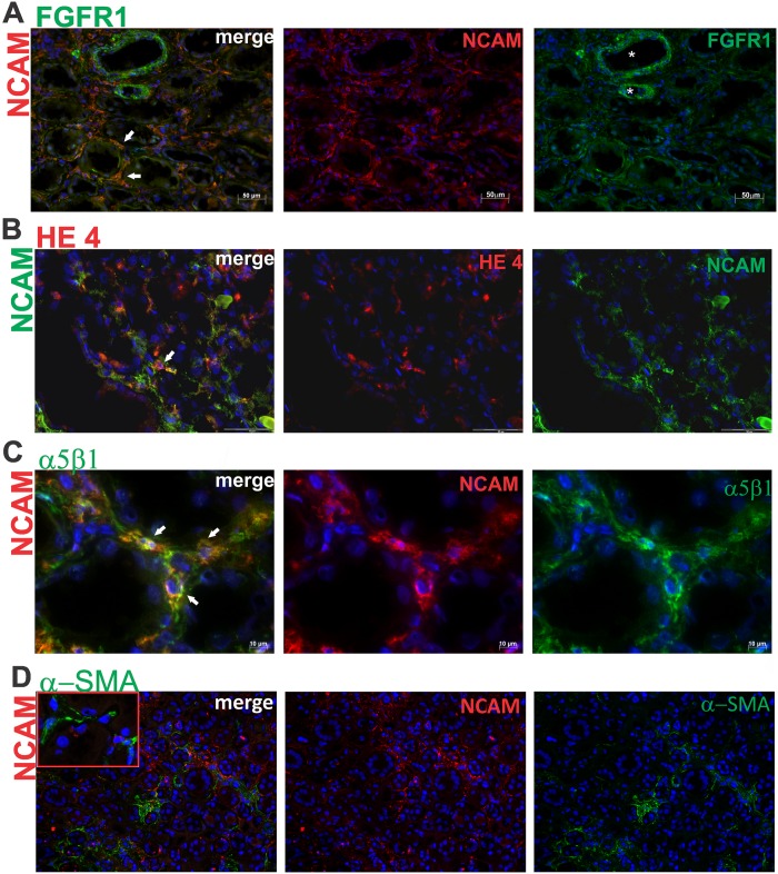
Fig 6. Expression of FGFR1, HE4 and α5β1 integrin on NCAM positive cells in incipient renal fibrosis.
(A) Double immunofluorescent labeling of NCAM and FGFR1; merge of these two markers clearly shows that all NCAM+ cells coexpressed FGFR1 (white arrows); diffuse NCAM expression on interstitial cells; strong FGFR1 expression on bold vessels (white stars) and diffuse expression on interstitial cells; x200. (B) Double immunofluorescent labeling of NCAM and HE4; merge of NCAM and HE4 revealed single cells coexpressing both markers (white arrow); x400. (C) Double immunofluorescence labeling of NCAM and α5β1; merge of these two markers clearly shows co-expression of NCAM and α5β1 on renal interstitial cells in area of incipient fibrosis (white arrows); x600. (D) Double immunofluorescence labeling of NCAM and αSMA; merge of these two markers showed no overlapping of NCAM and αSMA on renal interstitial cells in area of incipient fibrosis, although areas of NCAM+ and SMA+ interstitial cells are close to each other; x100. Staining techniques are described in detail under Material and Methods.

