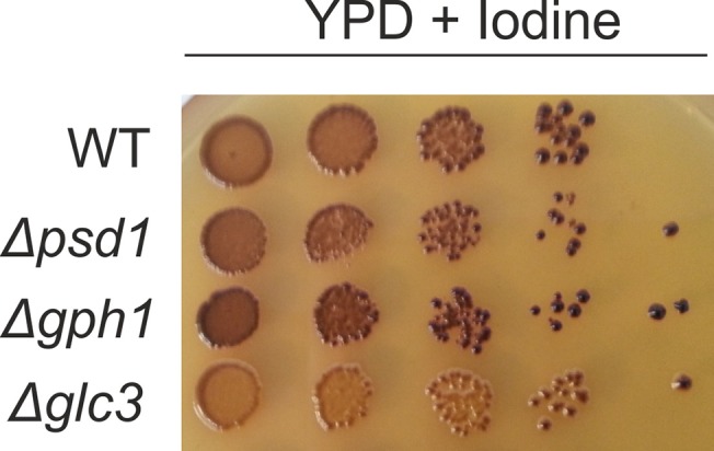
Fig 5. Glycogen content in the Δgph1, Δpsd1 and Δglc3 deletion mutants.

Strains as indicated were grown on YPD plates. Cell suspensions were spotted at dilutions 1, 1/10, 1/100, 1/1000, 1/10000 and incubated at 30°C for 48 h. Plates were exposed to iodine vapor for 10 min.
