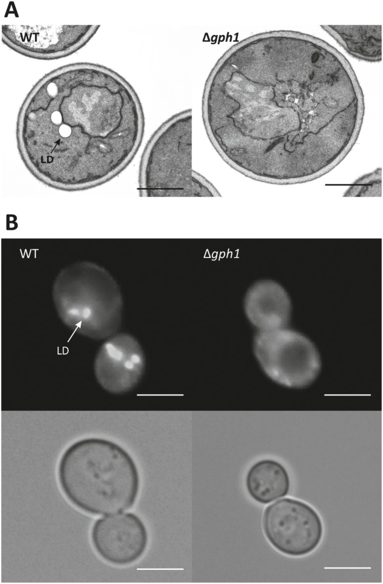
Fig 7. Number and size of lipid droplets are decreased in the Δgph1 deletion mutant.

(A) Transmission electron microscopy images of wild type BY4741 and Δgph1. (B) Nile Red staining and fluorescence microscopy of wild type BY4741 and Δgph1. LD: lipid droplets. Scale bar: 2 μm.
