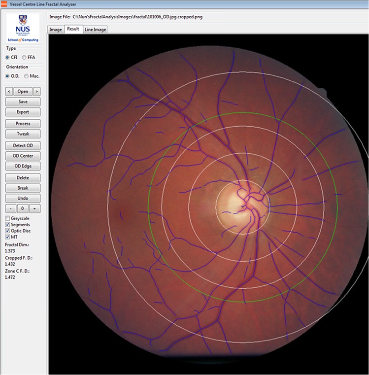
Fig 2. Retinal vascular fractal dimension measurement.

The upper image illustrates a retinal fundus image and skeletonized line tracing of an eye with a low fractal dimension and less complex (more rarefied) branching pattern; the lower retinal fundus image and skeletonized line tracing illustrates a higher fractal dimension and a more complex (dense) branching pattern.
