Figure 5.
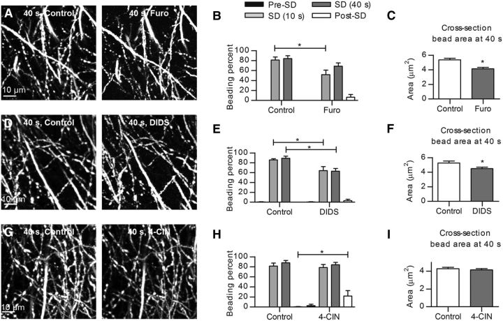
SD-induced dendritic beading is attenuated by separate inhibition of three different classes of cotransporters. A, Representative 2PLSM MIP images of dendritic beading at 40 s after SD onset elicited before (control) and 45 min after pretreatment with 1 mm furosemide. B, Beading before and after furosemide treatment shows significant difference at 10 s after SD onset (n = 6 slices, two-way RM-ANOVA and Sidak's post hoc test), post-SD quantified at 250 s. *p < 0.01. C, Quantification of the size of beads at 40 s after SD onset reveals significant reduction in the cross-section bead area in furosemide-containing aCSF (n = 36 beads in 6 slices, paired t test). *p < 0.001. D, Representative images of beaded dendrites at 40 s after SD onset in control condition and after 45 min of pretreatment with 300 μm DIDS. E, SD-induced beading was significantly reduced in DIDS-containing aCSF at 10 and 40 s after SD onset (n = 6 slices, two-way RM-ANOVA and Sidak's post hoc test), post-SD quantified at 250 s. *p < 0.001. F, The size of beads at 40 s after SD initiation in DIDS-containing aCSF was significantly decreased (n = 36 beads in 6 slices, paired t test). *p < 0.001. G, 2PLSM MIP images showing dendritic beads of similar sizes at 40 s after SD onset evoked in control condition and after 45 min of pretreatment with 0.5 mm 4-CIN. H, SD-induced dendritic beading percentage was unaffected by 4-CIN, whereas a slower recovery of beading was observed in 4-CIN-containing aCSF compared with control (n = 7 slices, two-way RM-ANOVA and Sidak's post hoc test), post-SD quantified at 250 s. *p < 0.01. I, Summary from 36 beads in 6 slices demonstrating no significant decrease in the size of beads at 40 s during SD after 4-CIN pretreatment (Wilcoxon's signed-rank test, p = 0.07).

