Figure 1.

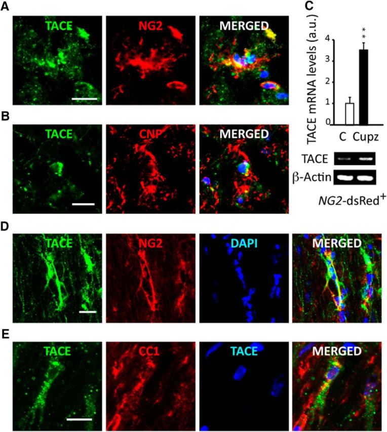
TACE is expressed by OL lineage cells in the white matter of subjects with MS and is upregulated following cuprizone-induced demyelination. A, B, Postmortem brain tissue samples from subjects with SPMS were used to characterize TACE expression in OL lineage cells in NAWM regions. Immunofluorescence analysis revealed TACE expression in OPs (A; NG2+ cells) and cells undergoing OL differentiation (B; CNP+ cells). C–E, TACE is upregulated in OL lineage cells in the SCWM following Cupz-induced demyelination. Adult NG2-dsRed mice were fed with Cupz or normal chow for 9 consecutive weeks. C, NG2-dsRed+ FAC-sorted cells were obtained from the SCWM of mice fed with Cupz or normal diet for 9 weeks, and TACE mRNA levels were analyzed by qPCR. D, E, Immunofluorescence analysis in the mouse SCWM after 9 weeks of Cupz treatment revealed TACE expression in OPs (D; NG2+) and cells undergoing OL differentiation (E; CC1+). Histograms are expressed as arbitrary units (a.u.) after β-actin normalization. Data from animal models are shown as mean ± SEM n = 4–6 brains for each time point. Scale bars, 10 μm. **p < 0.01 versus controls.
