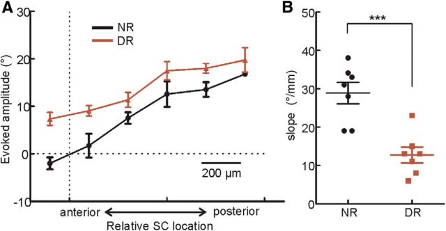
Figure 3.
Visual experience is required for the normal development of eye movement maps in the mouse SC. A, Horizontal amplitudes of evoked saccades in DR (brown) and NR (black) mice. The visual receptive fields of the superficial SC were mapped for some penetrations to determine the SC location that represented the vertical meridian, which was used to align maps from different mice. B, Eye movement maps in DR mice had a smaller slope along the anterior–posterior axis than in the NR mice (p < 0.001, t test). Each data point represents the slope from an individual animal. Pooled data were presented as mean ± SEM.

