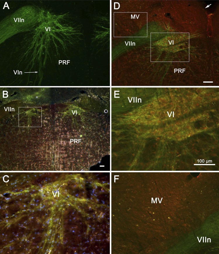
Fig. 1.
Histochemical demonstration of channelrhodopsin expression in motoneurons of the abducens nucleus (VI) of ChAT-ChR2 mice used in the optogenetic stimulation experiments. VI may be seen in these transverse sections (A–F) lying ventromedial to the genu of the facial nerve (VIIn). Note in A the wide extent of the dendritic arbors. Low (B and C)- and high-magnification views (D–F) of double-labeled tissue from 2 additional animals are shown. Anti-choline acetyltransferase (ChAT) marker is red, channelrhodopsin marker is green, and their overlap appears as a yellow-orange color. Like abducens motoneurons, cholinergic medial vestibular (MV) neurons show coexpression of the ChAT and channelrhodopsin in their somata but lack the motoneurons' dendritic expression of channelrhodopsin. In B–F, nuclei fluoresce blue. Boxes in B and D indicate location of higher magnification views. Arrow in D indicates the ventricular surface. All scale bars are 100 μm; scale bar in D also represents A, and scale bar in E also represents C and F. MV, medial vestibular nucleus; PRF, pontine reticular formation; VIn, abducens nerve.

