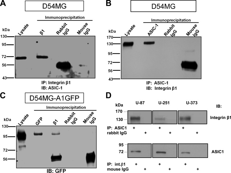
Fig. 1.
Physical association of acid-sensing ion channel (ASIC)-1 with integrin-β1 in glioma cells. A: cell lysates were immunoprecipitated (IP) with mouse anti-integrin-β1 (β1) antibody and immunoblotted (IB) with rabbit anti-ASIC-1. Immunoprecipitation of integrin-β1 pulled down ASIC-1 at ∼70 kDa. Lysate refers to total lysate (25 μg of protein) prepared from cells. B: immunoprecipitation of ASIC-1 using rabbit anti-ASIC-1 from D54MG cell lysates pulled down integrin-β1 detected by mouse anti-integrin-β1 at ∼130 kDa. C: in D54MG cells stably overexpressing GFP-tagged ASIC-1 (D54MG_A1GFP), immunoprecipitation of integrin-β1 pulled down GFP-ASIC-1 consistent with the expected size of GFP-tagged ASIC-1 at ∼100 kDa. Nonimmune rabbit IgG and mouse IgG did not immunoprecipitate any specific bands (n ≥ 3). D: ASIC-1 and integrin-β1 also coprecipitated from 3 additional glioma cell lines (U87, U251, and U373).

