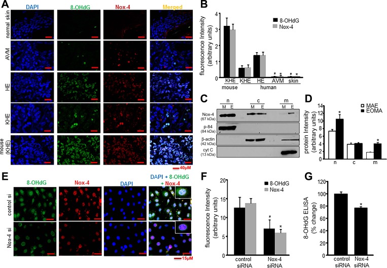
Fig. 3.
Nox-4-derived oxidants induce nuclear DNA oxidation. A: confocal images of immunofluorescence detection of 8-OHdG (1:75) and Nox-4 (1:100) in human tissue sections. DAPI was used to counterstain the nucleus. B: fluorescence intensity analysis (Olympus FV10-ASW software) showed significant differences in both 8-OHdG and Nox-4 detection between tumor-forming and non-tumor-forming ECs; *P < 0.05 for Nox-4 (HE vs. others) and #P < 0.05 for 8-OHdG (HE vs. others) of 5 determinant sections. C: Nox-4 localization in subcellular fractions of nucleus (n), cytosol (c), and mitochondrial membrane (m) was detected by Western blots in both cell types of EOMA (E) and murine aortic endothelial (MAE) (M), which was normalized against housekeeping proteins as described in materials and methods. D: representative band intensity was measured using ImageJ software. E: immunocytochemistry shows colocalization of 8-OHdG (1:100), Nox-4 (1:100), and DAPI (1:10,000) in Nox-4 knockdown EOMA cells. F: fluorescence intensity quantified using Olympus FV10-ASW software shows elevated levels of 8-OHdG and Nox-4 in tumor-forming vs. non-tumor-forming ECs. G: significant decrease in 8-OHdG levels measured by ELISA in EOMA cells after nox-4 siRNA transfection. Results are expressed as means ± SD; *P < 0.05. For tumor sections, n = 5 fields per section.

