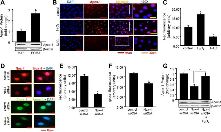
Fig. 4.
Nox-4-derived H2O2 induces expression of Apex-1. A: Western blot shows elevated Apex-1 protein expression in tumor-forming EOMA cells vs. non-tumor-forming MAE cells. B: H2O2-induced Apex-1 expression was measured by immunocytochemistry. Cells were challenged with H2O2 (250 μM) for 6 h and then treated with N-acetyl cysteine (NAC, 5 mM) for 12 h. C: quantification of fluorescence intensity for Apex-1 was analyzed. D: Apex-1 expression measured by immunocytochemistry was significantly reduced in Nox-4 knockdown cells. E and F: quantification of fluorescence intensity of both Nox-4 and Apex-1. G: rescue effect has been established by posttreatment of H2O2 after knocking down Nox-4. Results are expressed as means ± SD; *P < 0.05.

