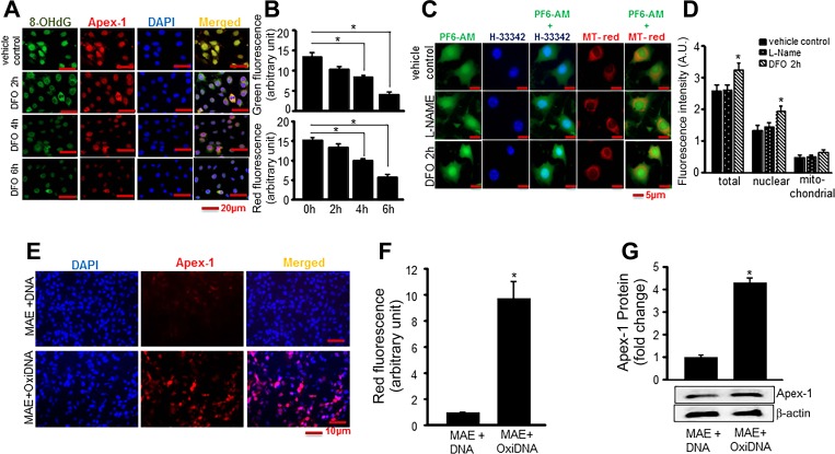
Fig. 5.
Nox-4-derived oxidants cause DNA oxidation that stimulates Apex-1 expression. A: accumulation of 8-OHdG in nuclei of EOMA cells was decreased in response to deferoxamine (DFO) treatment. EOMA cells were grown on coverslips. After 18 h, cells were treated with DFO (100 μM) for specified time points as indicated. Cells were fixed and stained with 8-OHdG (green), Apex-1 (red), and DAPI (blue). B: fluorescence intensity determines there was significant decrease of both Apex-1 and 8-OHdG expression with increasing time points. C: accumulation of H2O2 in presence of DFO was determined by a specific cell-permeable dye PF6-AM that interacts with intracellular H2O2 and generates a signal (green). Levels of colocalization were analyzed by nuclear (Hoechst 33342) and mitochondrial (MitoTracker Red) staining. l-NAME was used as inhibitor for peroxynitrite to confirm the specificity of PF6-AM. D: quantification of fluorescence intensity determines increased accumulation of H2O2 after 2 h of DFO treatment in total as well as nuclear compartments. E: images display oxidized DNA increased expression of Apex-1. Oxidative DNA was isolated from EOMA cells and transfected to MAE cells for 48 h as described in Methods. F: quantification of fluorescence intensity of Apex-1 in presence of oxidized DNA. G: Apex-1 protein expression in presence of oxidative DNA measured by Western blot analysis. Results are expressed as means ± SD; *P <0.05.

