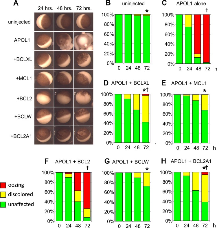
Fig. 1.
Toxic effects of apolipoprotein L1 (APOL1) expression in Xenopus oocytes are rescued or attenuated by coexpression of BCL2 family members. A: representative micrographs of oocytes recorded 24, 48, and 72 h post-injection of APOL1 cRNA (10 ng) without or with cRNA encoding the indicated individual BCL2 family polypeptides (50 ng), compared with uninjected oocyte controls (top row). Oocyte viability was graded with three toxicity categories: “unaffected” (resembling uninjected oocytes, i.e., complete rescue), “discolored” (with variably mottled or depigmented animal pole), and “oozing” or “blebbing” (with loss of physical integrity). The colored bars in the histograms correspond to the key in the bottom left: unaffected (green), discolored (yellow), and oozing (red). B and C: uninjected oocytes and oocytes injected with 10 ng APOL1 cRNA were imaged and graded at time t = 24, 48, and 72 h post-injection. C–H: oocytes coinjected with 10 ng APOL1 cRNA and 50 ng cRNA encoding the indicated BCL2 family polypeptides were imaged and graded at t = 24, 48, and 72 h post-injection. n = 40 in each group, made up of 10 oocytes from each of 4 frogs. Pairwise statistical differences between groups at 72 h post-injection were determined by ANOVA of ranks with a Tukey post hoc test. *P < 0.05 vs. oocytes expressing APOL1 alone. †P < 0.05 vs. uninjected oocytes.

