Fig. 2.
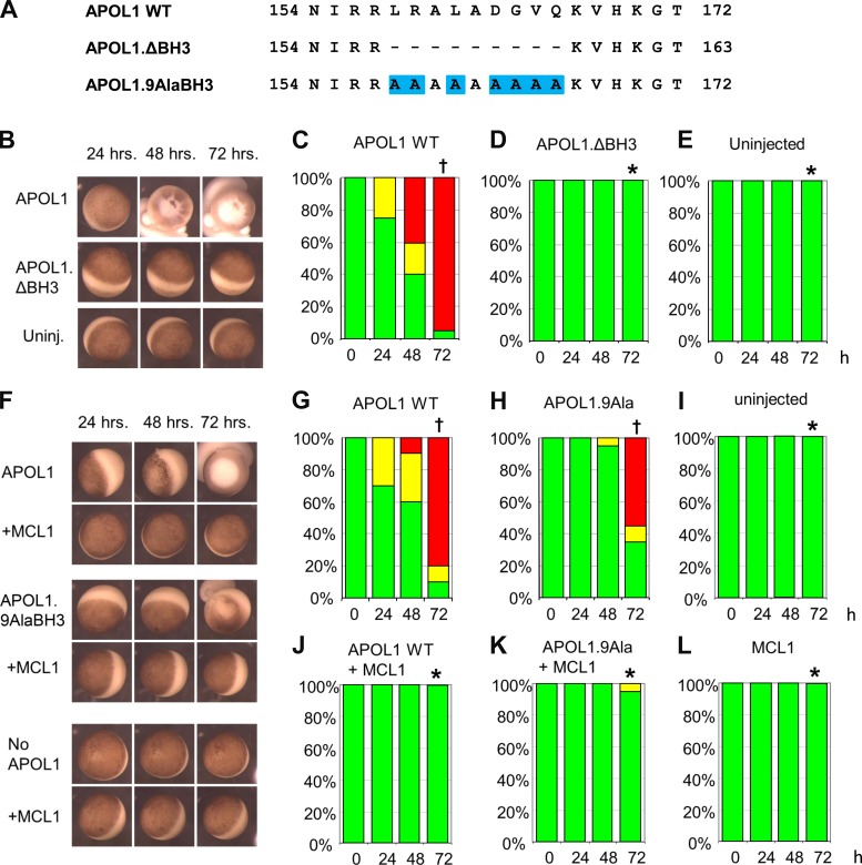
APOL1 BH3 domain deletion (APOL1.ΔBH3) prevents APOL1-associated oocyte toxicity, whereas BH3 Ala(9) substitution (APOL1.9AlaBH3) only minimally reduces oocyte toxicity but does not inhibit rescue of toxicity by MCL1. A: aligned BH3 domain sequences of wild-type (WT) APOL1 and of BH3 mutants, bracketed by amino acid sequence positions (numbers). Mutated residues are highlighted in blue. B: representative oocytes imaged at the indicated times post-injection with cRNA (10 ng) encoding APOL1 or APOL1 ΔBH3, compared with uninjected oocytes (bottom row). C–E: oocyte morphology category histograms at 24, 48, and 72 h post-injection of cRNA (10 ng) encoding APOL1 (C) or APOL1.ΔBH3 (D), compared with uninjected oocytes (E). n = 20 in each group (10 oocytes from each of 2 frogs). F: representative oocytes imaged at the indicated times post-injection with cRNA (10 ng) encoding APOL1 WT or APOL1.9AlaBH3, without or with coinjected MCL1 cRNA (50 ng), and compared with uninjected oocytes or oocytes injected with MCL1 alone (bottom). G–L: oocyte morphology histograms at 24, 48, and 72 h post-injection of the indicated cRNAs. n = 20 in each group (10 oocytes from each of 2 frogs). Statistical differences among groups at 72 h was determined as described for Fig. 1. *P < 0.05 vs. oocytes expressing APOL1 alone. †P < 0.05 vs. uninjected oocytes.

