Fig. 7.
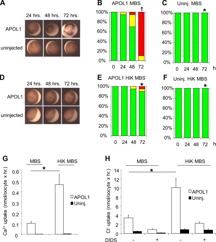
Oocyte exposure to high extracellular [K+] for 3 days post-injection of APOL1 cRNA attenuates morphological toxicity and increases ion fluxes in surviving oocytes. A: representative uninjected oocytes and oocytes injected with 10 ng APOL1 cRNA and maintained in MBS for the indicated times. B: oocyte morphology histograms at 24, 48, and 72 h post-injection of APOL1 cRNA with subsequent maintenance in MBS. C: oocyte morphology histograms for uninjected oocytes maintained in MBS for 24, 48, and 72 h. D: representative uninjected oocytes and oocytes injected with 10 ng APOL1 cRNA after maintenance in high K+ (HiK) MBS for the indicated times. E: oocyte morphology histograms at 24, 48, and 72 h post-injection of APOL1 cRNA, with maintenance in HiK MBS. F: oocyte morphology histograms for uninjected oocytes maintained in HiK MBS for 24, 48, and 72 h. n = 10 oocytes in each group in E and F. *P < 0.05 vs. oocytes expressing APOL1 alone. †P < 0.05 vs. uninjected oocytes. One of six similar experiments is shown. G and H: oocytes injected with APOL1 cRNA 72 h previously with subsequent maintenance in HiK MBS exhibit higher 45Ca2+ uptake (G) and higher DIDS-sensitive 36Cl− uptake (H) than oocytes maintained in MBS. Solid bars, uninjected oocytes. Values are means ± SE; n = 10 oocytes in each group. *P < 0.05.

