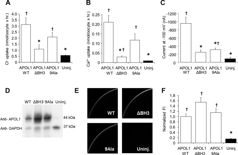
Fig. 9.
APOL1-associated oocyte uptakes of Ca2+ and Cl− are nearly abolished by BH3 domain deletion, but not by Ala substitution. Oocytes maintained 72 h in HiK MBS following injection with 5 ng cRNA encoding APOL1 WT, APOL1 ΔBH3, or APOL1 9Ala were maintained compared with uninjected oocytes in assays of 45Ca2+ uptake (n = 20 from 2 frogs; A), 36Cl− uptake (n = 20 from 2 frogs; B), and current at −100 mV as measured by two-electrode voltage clamp (n = 10 oocytes per group; C). D: immunoblot of detergent lysates (20 mg protein per lane) from oocytes expressing APOL1.WT, APOL1.ΔBH3, or APOL1.9Ala compared with lysate from uninjected oocytes. Simultaneous GAPDH probing of the same blot confirmed equal loading of lanes. One of two similar experiments with indistinguishable results is shown. E: representative median intensity images of whole mount confocal immunofluorescence micrographs of oocytes injected 72 h previously with the same WT and mutant APOL1 cRNAs, then maintained in HiK MBS prior to fixation and immunostaining with anti-APOL1 antibody. F: normalized mean fluorescence intensity values from oocytes treated as in D. Values are means ± SE; n = 20 (10 oocytes from each of 2 frogs). *P < 0.05 vs. oocytes expressing APOL1 alone. †P < 0.05 vs. uninjected oocytes.

