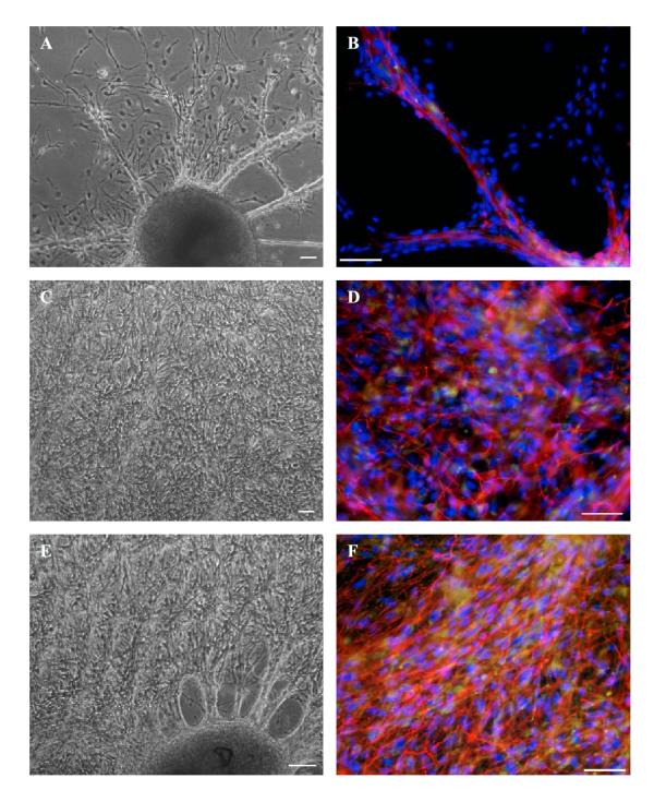
Figure 2.
Neuronal differentiation of monkey ES cells under the influence of monkey brain tissue crude extracts. Images were taken before (live) and after (fixed) immunostaining for TH (green) and TujIII (red). Co-expression of TH and TujIII appears in yellow. Cell nuclei were counterstained with DAPI (blue). A and B, without extracts; C and D, with cortical extracts; E and F, with striatal extracts. Scale bars are equivalent to 50 μm.

