Fig. 1.
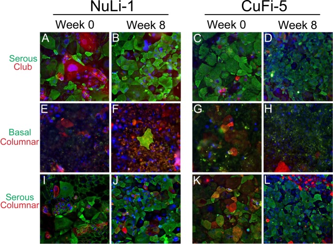
NuLi-1 and CuFi-5 cells display a serous cell phenotype at week 8 of air-liquid interface (ALI) culture. NuLi-1 and CuFi-5 cells display a predominantly pseudostratified serous cell phenotype according to lectin analysis, and apical cell surfaces of these cells preferentially bind lectins that associate with serous cell types, such as goblet and club cells. Cells were analyzed at initial shift to ALI (week 0) and at week 8 post-ALI. Cell type-specific sugar-binding lectins were used to phenotype cells. Lectins and the cells they stain were as follows: jack fruit lectin (jacalin/A. integrifolia), serous cells; MPL lectin (M. pomifera), club cells; peanut lectin (PNA, A. hypogaea), columnar cells; BSI-B4 lectin (B. simplicifolia), basal cells. A–D: cells labeled with jacalin (green, serous) and MPL (red, club). E–H: cells stained with BSI-B4 (green, basal) and PNA (red, columnar). I–L: cells stained with jacalin (green, serous) and PNA (red, columnar). Nuclei are stained with DAPI (blue). Not all cells stained at similar intensities, most likely due to specific characteristics of each cell type, such as abundance of sugars on the cell surface. Since at week 8 not all cells had comparable staining intensity, there may be multiple cell types, such as serous pseudostratified and columnar cells, within the cultures. Also note the various phenotypes and sizes of cells grown with submerged culture conditions at week 0 (A/C, E/G, and I/K) compared with week 8 (B/D, F/H, and J/L) post-ALI, where cells appear more disorganized at week 0 and more regular at week 8. Cells at passages 5–17 were used in this analysis. Magnification ×20 for all images.

