Fig. 4.
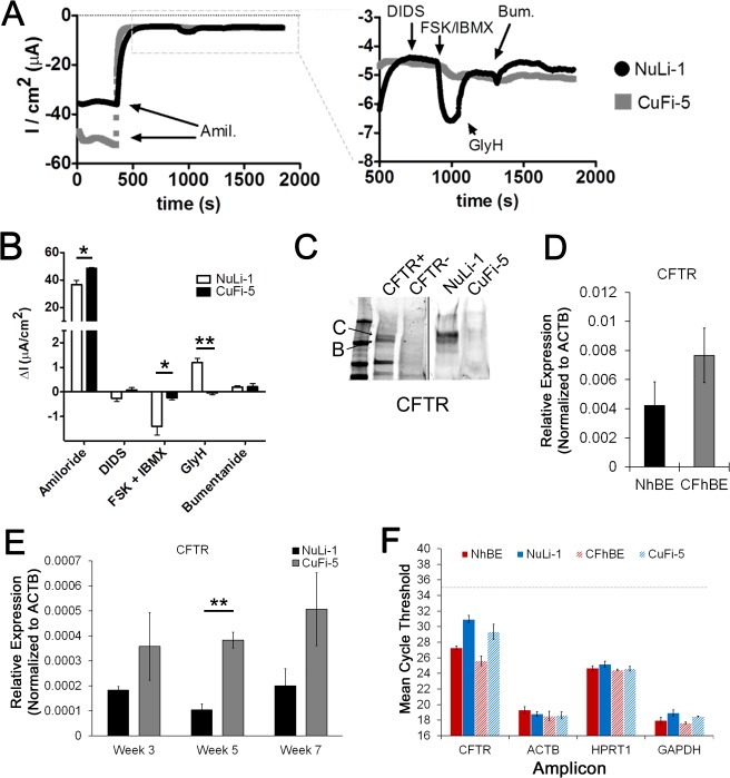
Ussing chamber traces of NuLi-1 vs. CuFi-5 cells reflect low CFTR transcript expression at week 7 of ALI culture. A: representative transepithelial recordings of NuLi-1 and CuFi-5 filters at week 7 post-ALI, recorded simultaneously. Left: entire trace. Addition of 10 μM amiloride (Amil) to the apical chamber resulted in a rapid decrease in negative current, consistent with a reduction in ENaC-mediated Na+ current. Right: magnification of the stabilized current after addition of amiloride. Addition of 10 μM DIDS, to block Ca2+-activated Cl− channels, to the apical side did not change current in either filter. CFTR agonists, 10 μM FSK and 100 μM IBMX, added to the basolateral side increased negative current in NuLi-1, but not CuFi-5, cells, consistent with activation of membrane-localized CFTR. FSK/IBMX-stimulated current was inhibited by addition of 20 μM GlyH to the apical chamber. Addition of 100 μM bumetanide (Bum) did not induce further inhibition of the current. B: week 7 results complementing trace in A. CuFi-5 cells were unresponsive to CFTR agonists and antagonists but displayed increased amiloride-sensitive current at baseline, characteristic of primary CF epithelia (15, 60). Values are means ± SE (n = 3–4 filters for each time point). C: representative Western blot of CFTR expression in NuLi-1 and CuFi-5 cells. Exogenous expression of wild-type CFTR in HeLa cells was used for antibody control (CFTR+, contrast enhanced for clarity). CFTR bands B and C are designated as such. D: gene expression analysis of CFTR in primary hBE cells grown on permeable supports. Exression levels of CFTR gene trends higher in CFhBE than NhBE cells. E: gene expression analysis of CFTR in NuLi-1 and CuFi-5 cell model indicates that gene expression stably trends higher over time in CuFi-5 than NuLi-1 cells. **P ≤ 0.01 (by 1-way ANOVA with Bonferroni's correction). F: mean CT values of CFTR and 3 housekeeping genes for reference and comparison. Expression trends and data corroborate electrophysiological properties of the cells (A and B), where Cl− currents are ≥10-fold lower than published expected Cl− currents for primary ciliated airway epithelial cells (15, 60).

