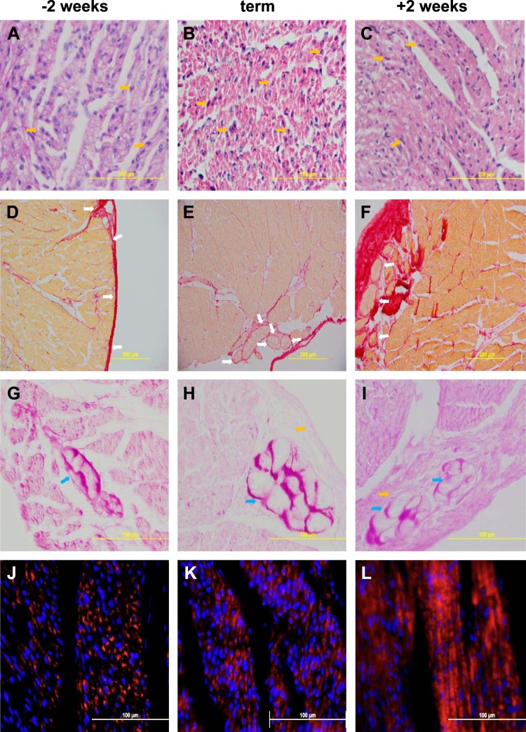
Fig. 4.
Representative illustrations of the histological changes in the heart, occurring from 2 wk before term (A, D, G, J), to term (B, E, H, K) and to 2 wk postnatally (C, F, I, L). Top row, A–C: hematoxylin and eosin staining illustrating increased density of the myocardium; dividing nuclei are seen at all ages (examples shown yellow arrows). 2nd row, D–F: picrosirius red staining for collagen (red), showing increased density of the myocardium and increased collagen (red staining); Purkinje fibers in the collagen can be seen at all ages (examples shown with white arrows), although Purkinje fibers are more difficult to identify in the younger fetus because of the relatively low collagen deposition around them. 3rd row, G–I: periodic acid-Schiff staining illustrating Purkinje fibers (blue arrows) containing plentiful glycogen (purple); collagen surrounds them, orange arrows (H, I). 4th row, J–L: COX IV (red) staining in mitochondria and nuclear DNA stained with Hoechst 33342 (blue) show increased mitochondrial density and organization with maturation.

