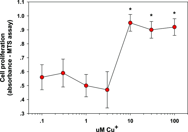
Fig. (1).
Stimulation of Fibroblast proliferation by Copper. Fibroblasts were exposed to 0–100 µM copper for 24 hr. The cells were examined for proliferation by MTS assay. Effects are represented as absorbance units; * = p < 0.05, relative to control (0 Cu2+). Error bars represent standard deviation, n = 4. Data taken from [26].

