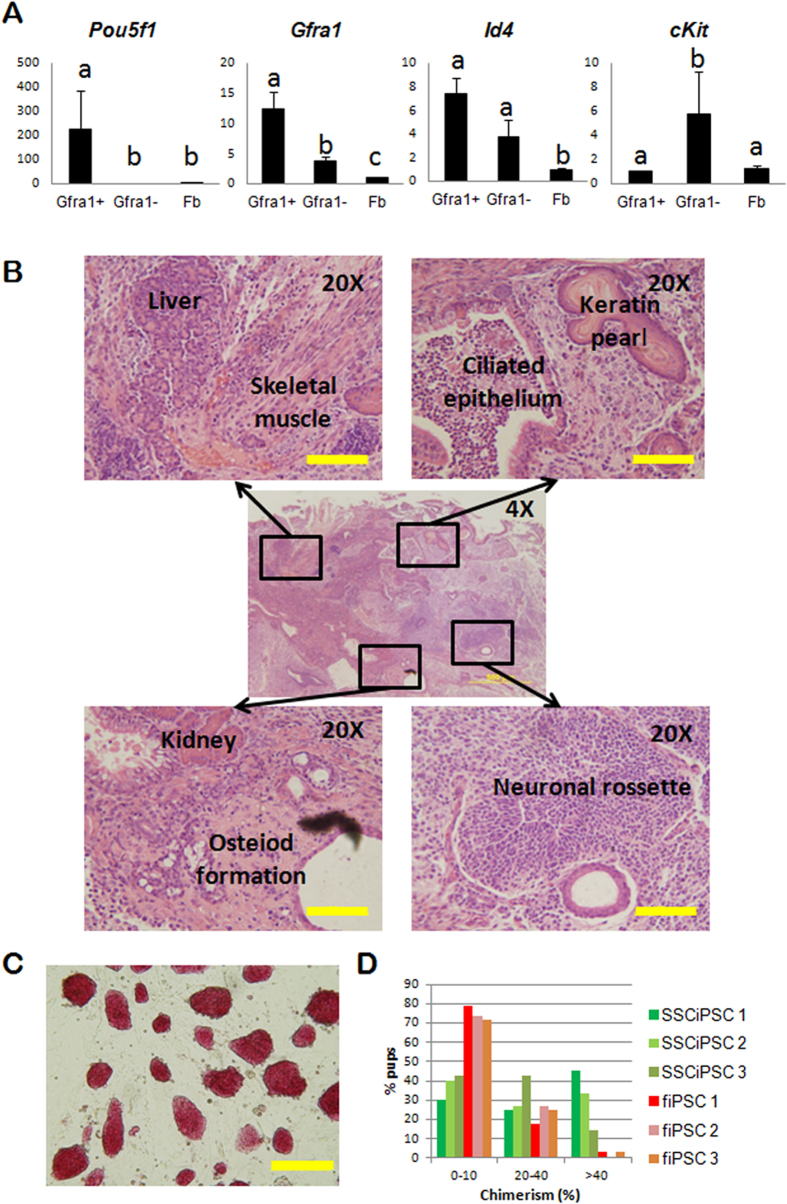
Figure 1. Reprogramming and characterization of iPSC derived from SSC.
(A) Relative mRNA abundance determined by qPCR of three genes expressed in SSC (Pou5f1, Gfra1 and Id4) and a marker of differentiated spermatogonia (cKit) in the testicular population sorted for Gfra1 (Gfra1+), unsorted (Gfa1−), and somatic fibroblast controls. Different letters indicate significant differences between treatments based on ANOVA (p < 0.05). (B) H&E histological section of a subcutaneous teratoma generated by SSCiPSC injection into immunocompromised mouse. The central image shows a 4X overview of the teratoma [Scale bar: 500 μM]. Four areas are amplified (20X) in the side images showing derivatives from the three germ layers [Scale bar: 100 M]. (C) Representative 10X light microscope image of SSCiPSC colony morphology after alkaline phosphatase staining [Scale bar: 200 μM]. (D) Percentage of skin chimerism in pups derived from 3 different SSCiPSC lines (green bars) or fiPSC (red-brown bars).

