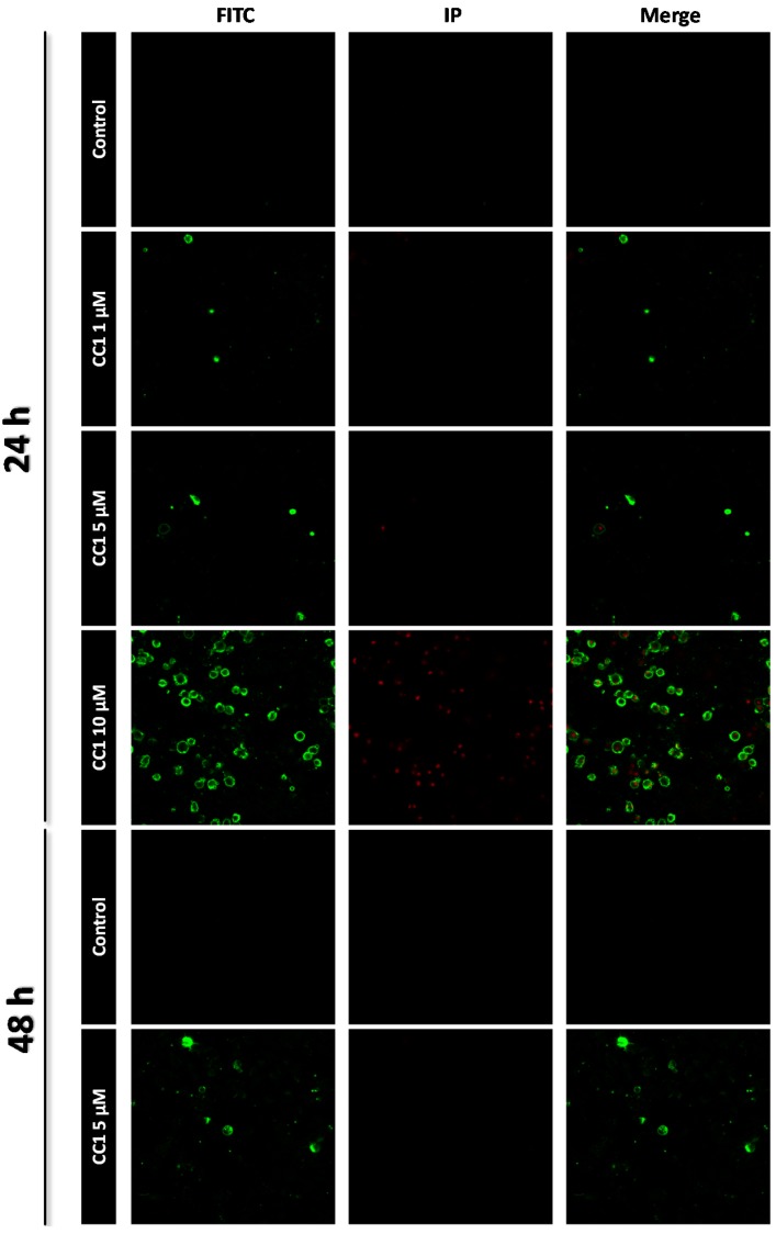
Figure 2.
Apoptosis detection by confocal microscopy after 24 h and 48 h treatments with 1, 5 and 10 μM crambescin C1 (CC1). Representative photos of control and treated cells are shown. Fluorescein isothiocyanate (FITC) was used for phosphatidylserine translocation detection (green) and propidium iodide (IP) was used for nuclei staining of death cells (red).

