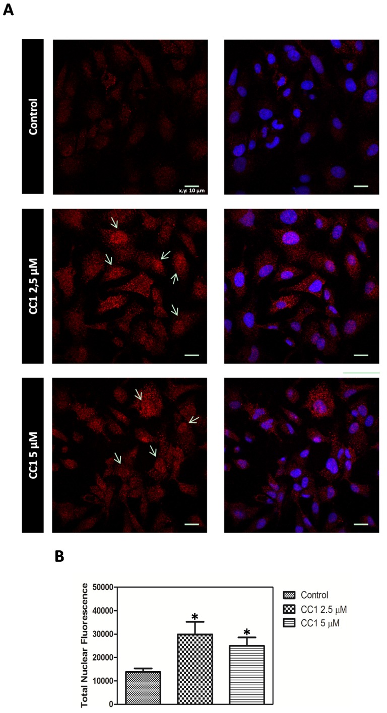
Figure 7.
(A) MT-1, -2 detection by confocal microscopy in control and HepG2 cells treated with 2.5 μM and 5 μM crambescin C1 (CC1) for 12 h. Representative photos of control and treated cells are shown. Hoechst 33258 was used for nuclei counterstaining (blue) and quantification of nuclear metallothioneins (MTs). Arrows: MTs translocation to the nucleus in treated cells; (B) Quantification of the variations caused by CC1 in the levels of nuclear MTs.

