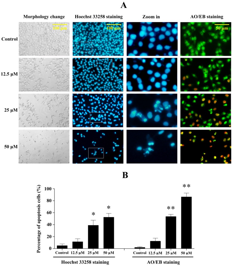
Figure 6.
Penicitrinine A induced significant apoptotic morphological changes. (A) After exposed to 12.5, 25 and 50 μM penicitrinine A for 24 h, A-375 cells were stained by Hoeschest 33258 or AO/EB. Photos were taken under an inverted fluorescence microscope; (B) Quantification of the proportion of apoptotic cells detected by Hoechst 33258 staining and AO/EB staining. The values (means ± SD, n = 3) differed significantly (* p < 0.05; ** p < 0.01). The third column in Figure A was amplified from the second column for ten fold.

