
Figure 16.
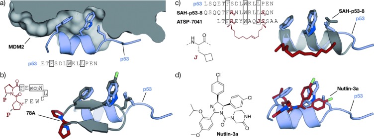
MDM2–p53 interaction: a) Crystal structures of MDM2 (gray) with the transactivation domain of p53 (blue, PDB 1YCR).[347] b) Superimposed crystal structures of p53 (blue, PDB 1YCR) and cyclic β-hairpin peptide 78A (gray/red, PDB 2AXI). The d-Pro-l-Pro (p-P) cross-link is highlighted in red.[115] c) Sequences of stapled peptides (left). Superimposed crystal structures (right) of p53 (blue, PDB 1YCR) and SAH-p53-8 (gray/red, PDB 3V3B). The cross-link is highlighted in red (side chains of amino acids in boxes are shown explicitly in the crystal structures).[355] d) Superimposed crystal structures of p53 (blue, PDB 1YCR) and Nutlin-3a (red, PDB 4HG7).[356] All superimposed structures were obtained from structures of complexes with MDM2 or MDMX.