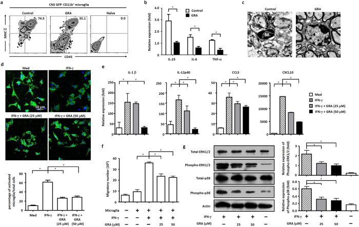
Figure 4. GRA inhibits microglia activation through suppression of MAPK signal pathway.
(a) Flow cytometry analysis of activation markers (CD45 and MHC ΙΙ) on gated GFP−CD11b+ microglia isolated from the CNS of naïve, control and GRA treatment GFP-chimeric mice assessed on 15 day post immunization. Data are representative of four mice per group. (b) qPCR analysis of the indicated genes in FACS-sorted GPF−CD11b+ microglia isolated from naïve, control and GRA treatment GFP-chimeric EAE mice on 15 day post immunization. (c) TEM analyses of microglia status in spinal cords of day 15 control and GRA-treated EAE mice. (d) Representative images of primary microglia cultured with medium alone or treated with IFN-γ (100 ng/ml) or IFN-γ (100 ng/ml) plus GRA at the indicated concentrations, and the percentage of activated microglia with amoeboid shape normalized to total microglia (CD11b+). CD11b, green; DAPI, blue. *P < 0.05. (e) Primary microglia were cultured with medium alone or treated with IFN-γ (100 ng/ml) or IFN-γ (100 ng/ml) plus GRA at the indicated concentrations. Quantification of mRNA abundance for IL-1β, IL-12p40, CCL5 and CXCL10. (f) Culture supernatants were collected and cocultured with DLN cells isolated from day 15 EAE mice. The chemotactic ability was analyzed by counting the amount of migratory cells in the lower chamber. (g) Protein extracts from cultured microglia for 48 h were analyzed by western blot for expression of phosphorylated ERK1/2 and p38. Quantification results were shown on the right. Data are representative of three independent experiments. *P < 0.05.

