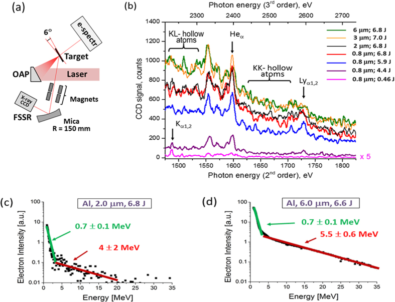
Figure 2. Scheme of observation and results.
(a) Experimental set up. The laser beam is focused by an off-axis parabola almost perpendicular to the surface of foil and heats the resulting plasma. The produced plasma generates X-ray emission, which was measured by X-ray high luminosity spectrometer with high spectral resolution placed at 45° to the target surface. Electron spectra were measured by electron spectrometer placed from rear side of plasma perpendicular to the target surface; (b) Single shot, spatially- and temporally averaged Al ions K-shell spectra (raw data) emitted from foil targets with different thickness and laser energies (Intensity of spectra obtained by irradiation of laser pulse with energy 0.46 J is multiplied by factor 5); (c,d) Experimentally measured electron energy distribution in the case of irradiation of 2 and 6 μm thickness Al foils by maximum laser intensity of 1021 W/cm2. The increase of electron temperature of the hot electrons Te,hot with the thickness of Al target is clearly observed.

