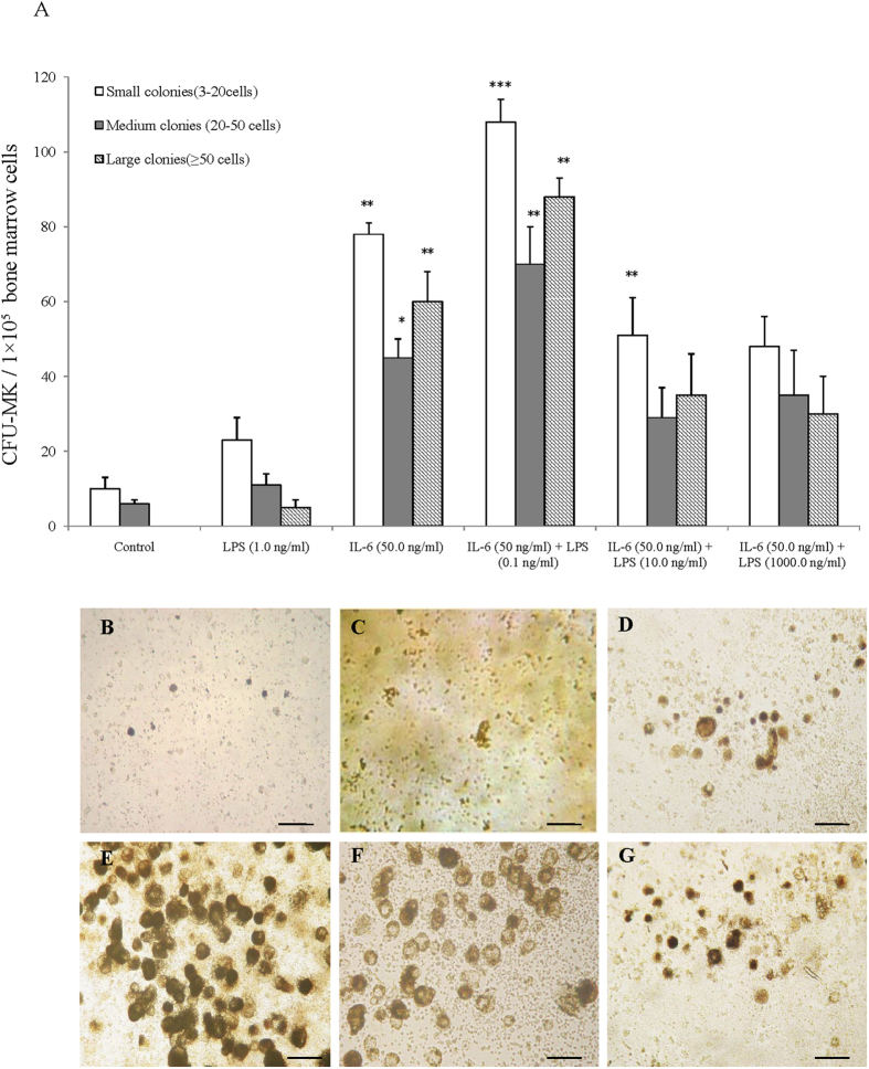
Figure 3. Effect of LPS alone or with IL-6 on CFU-MKs.
(A) Number of CFU-MKs in different mouse groups after incubation with LPS alone or IL-6 with LPS. Colonies classified according to their size were evaluated by inverted light microscopy. Experiments were performed in triplicate in four assays. Significant results compared with PBS are based on *P < 0.05 or **P < 0.01. (B) Bone marrow cells from TPO-pretreated mice were isolated by a discontinuous BSA density gradient and grown in a liquid serum-free medium in LPS alone or with IL-6 for 3 d. Cells were stained for acetylcholinesterase activity and phase contrast morphology of megakaryocyte cells. Scale bar, 10 μm.

