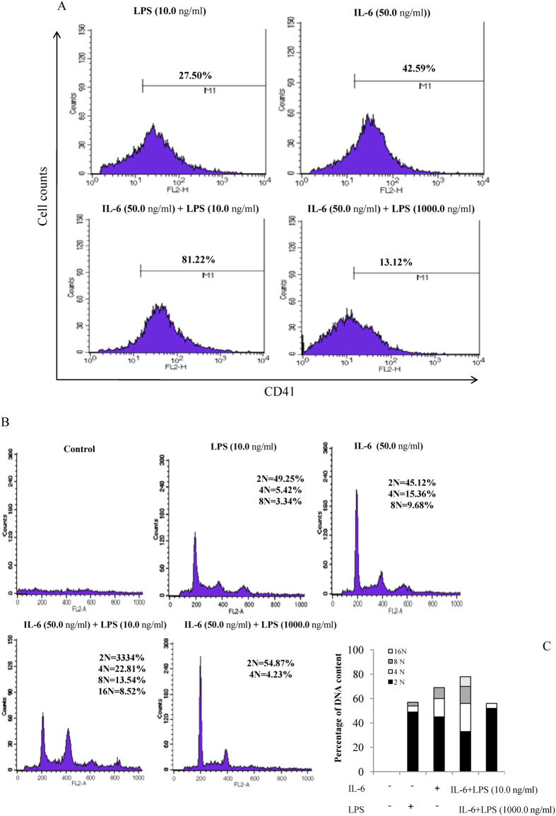
Figure 4. Effects of LPS alone or with IL-6 on CD41 expression and polyploidization.
(A) Bone marrow cells were grown in a liquid serum-free medium supplemented with LPS or IL-6 alone or together for 3 d. Megakaryocytes were stained with FITC-conjugated anti-mouse CD41 monoclonal antibody and analyzed by FACS with large forward-scatter and side-scatter properties. The experiment was repeated three times, and similar results were obtained. (B) Bone marrow cells from TPO-pretreated mice were isolated on a discontinuous BSA density gradient and grown in a liquid serum-free medium in the presence of IL-6 or LPS alone or together for 3 d. The flow cytometric analysis of the DNA content of CD41-positive cells was examined by PI staining. The experiment was repeated three times, and similar results were obtained. (C) The histogram plots of PI staining were calculated from the percentage of DNA content.

