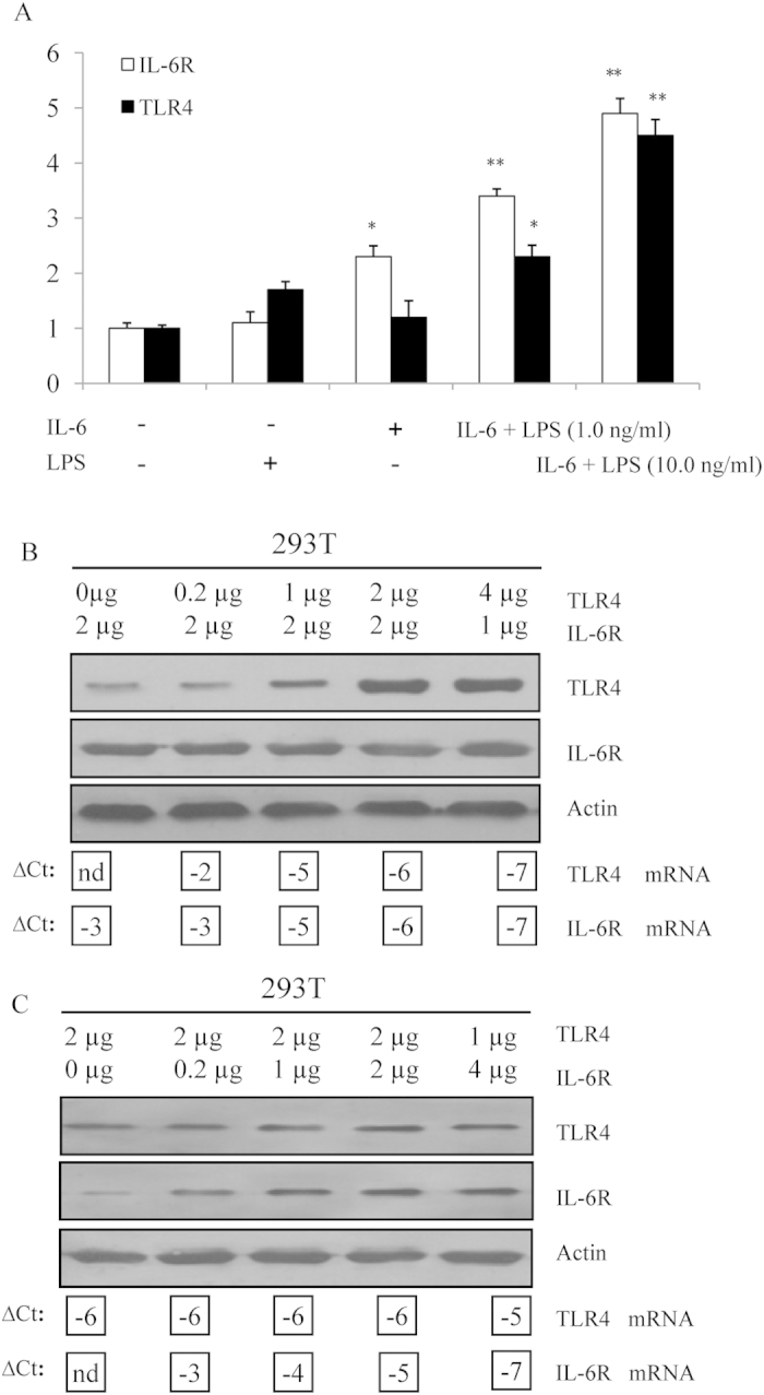
Figure 6. Dose-response relationships for TLR4 expression with IL-6R expression.

(A) Alteration in the expression of the TLR4 and IL-6R genes. Total RNA was extracted from Dami cells treated with LPS alone, IL-6 alone or LPS plus IL-6 for 48 h. The relative expression levels of TLR4 and IL-6R in the treated Dami cells were compared with those in the untreated Dami cells. All measurements show the expression relative to the expression levels of β-actin. The values are based on three independent experiments with triplicate measurements for each. **P < 0.01; ***P < 0.001. (B) 293T cells were transiently co-transfected with varying amounts of plasmid DNA encoding TLR4 and IL-6R. TLR4, IL-6R, and actin proteins were detected by immunoblot analysis. The mRNA expression of TLR4 and IL-6R was determined by Q-PCR; ∆CT values for TLR4 and IL-6R are shown below the corresponding lanes of the immunoblots. The amount of TLR4 DNA was kept constant, whereas the amount of IL-6R DNA was varied. (C) TLR4 DNA was varied, and the amount of IL-6R DNA was kept constant.
