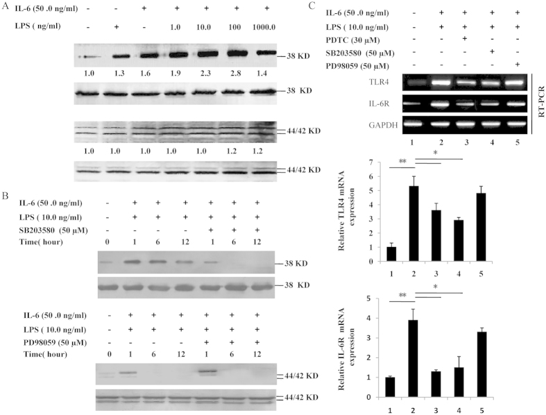
Figure 7. Phosphorylation of p38 MAPK and ERK1/2 and regulation of TLR4 and IL-6R mRNA expression.
(A) Dami cells were treated with medium alone or varying concentrations of LPS (1.0-1000.0 ng/ml) and IL-6 (50.0 ng/ml) for 24 h. The levels of phosphorylation of p38 MAPK and ERK1/2 were determined by western blot using the specific antibodies P-p38 MAPK and P-ERK1/2, respectively. Equal loading in the lanes was evaluated by probing with the T-p38 MAPK and T-ERK1/2 antibodies. The value under each sample indicates the fold change of the protein level relative to that of the control. (B) The LPS-induced activation of ERK and p38 kinase in Dami cells was inhibited by specific inhibitors. Dami cells were pretreated with PD98059 or SB203580 at 50 μM for 30 min before stimulation with LPS and IL-6. The corresponding western blots for the total levels of this kinase are labeled T-ERK1/2 and T-p38 MAPK, and the control was used to ensure equal loading of the proteins. (C) LPS-induced TLR4 and IL-6R mRNA expression was inhibited by PD98059, SB203580 or PDTC. Dami cells were pretreated with or without PD98059, SB203580 or PDTC for 30 min and then stimulated with LPS plus IL-6 for 24 h. The relative TLR4 and IL-6R mRNA expression present in unstimulated cells is expressed as 100%. Similar results were observed in three independent experiments.

